Shoulder Tendon Anatomy Diagram - Anatomy Of The Human Shoulder Joint - Along with muscles and tendons, they are a main source of stability for the shoulder.
byAdmin-
0
Shoulder Tendon Anatomy Diagram - Anatomy Of The Human Shoulder Joint - Along with muscles and tendons, they are a main source of stability for the shoulder.. The labrum also serves as the attachment of a major tendon in the shoulder, the biceps tendon. Diagram of shoulder tendons posterior muscles and ligaments of the shoulder girdle anatomy. You can see it enclosing the glenohumeral joint okay! Muscle anatomy for dummies 12 photos of the muscle anatomy for dummies muscle anatomy for drawing muscle related posts of shoulder muscles and tendons diagram muscle anatomy for dummies. Robin smithuis and henk jan van der woude.
The labrum also serves as the attachment of a major tendon in the shoulder, the biceps tendon. Draw labelled diagram showing the relations of shoulder joint. The shoulder anatomy provides mobility but leads to a relatively unstable joint, prone to subluxation and dislocation 2. Shoulder radiology & anatomy at usuhs.mil. Robin smithuis and henk jan van der woude.
Rotator Cuff Tears Frequently Asked Questions Orthoinfo Aaos from orthoinfo.aaos.org Prevents inferior translation and external rotation in the abducted shoulder, and provides stability to the long head of the biceps tendon (neer cs ii, corr 1992;280:182). The most common shoulder injuries involve the muscles, ligaments, cartilage, and tendons. Normal anatomy, variants and checklist. Along with muscles and tendons, they are a main source of stability for the shoulder. Draw labelled diagram showing the relations of shoulder joint. Shoulder muscles and shoulder tendons. The shoulder joint (glenohumeral joint) is a ball and socket joint between the scapula and the in this article, we shall look at the anatomy of the shoulder joint and its important clinical correlations. Three bones come together at the shoulder joint.
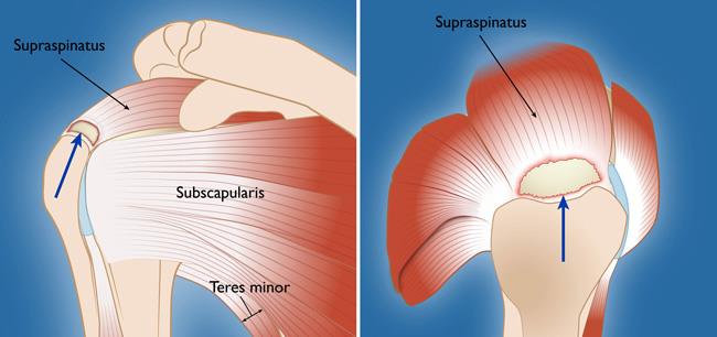
Prevents inferior translation and external rotation in the abducted shoulder, and provides stability to the long head of the biceps tendon (neer cs ii, corr 1992;280:182).
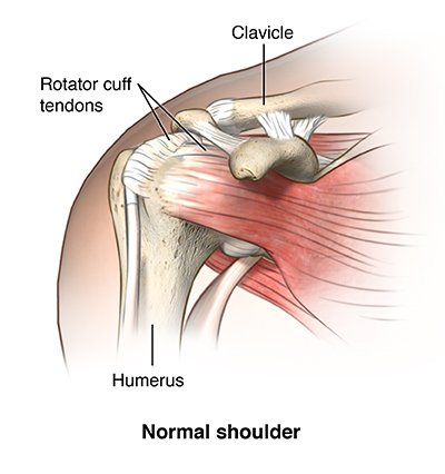
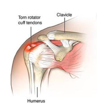
The shoulder anatomy includes the anterior deltoid, lateral deltoid, posterior deltoid, as well as the 4 rotator cuff muscles. Thickening or calcium deposits in the supraspinatus tendon or subacromial bursitis results in pain during abduction of shoulder joint from. Draw labelled diagram showing the relations of shoulder joint. Muscles allow us to move by pulling on bones. Normal anatomy, variants and checklist. An image depicting shoulder anatomy can be seen below. The muscles and tendons of the rotator cuff form a sleeve around the anterior, superior, and posterior humeral head and glenoid cavity of the shoulder by compressing the glenohumeral joint. Upper limb trauma programme injuries. Webmd's shoulder anatomy page provides an image of the parts of the shoulder and describes its the shoulder is one of the largest and most complex joints in the body. Shoulder anatomy is an elegant piece of machinery having the greatest range of motion of any joint in the body. The shoulder joint is the connection between the chest and the upper extremity. It can help you understand our world more detailed and specific. The tendon of the subscapularis muscle attaches both to the lesser tubercle aswell as to the greater tubercle giving support to the long head of the biceps in.
Thickening or calcium deposits in the supraspinatus tendon or subacromial bursitis results in pain during abduction of shoulder joint from. Shoulder radiology & anatomy at usuhs.mil. Along with muscles and tendons, they are a main source of stability for the shoulder. The long head and the short head. This diagram with labels depicts and explains the details of shoulder tendons and muscles.
Rotator Cuff Injury Cedars Sinai from api.kramesstaywell.com The shoulder muscles bridge the transitions from the torso into the head/neck area and into the upper extremities of the arms and hands. It can help you understand our world more detailed and specific. Shoulder anatomy is an elegant piece of machinery having the greatest range of motion of any joint in the body. Biceps and triceps tendon rupture. Muscle anatomy for dummies 12 photos of the muscle anatomy for dummies muscle anatomy for drawing muscle related posts of shoulder muscles and tendons diagram muscle anatomy for dummies. Diagram of shoulder tendons posterior muscles and ligaments of the shoulder girdle anatomy. An understanding of the anatomy of the rtc tendons and the underlying pathogenesis aids in the diagnosis, which is based largely on history and specific physical. Draw labelled diagram showing the relations of shoulder joint.
Draw labelled diagram showing the relations of shoulder joint.
For that reason, and because of the dexterity of the shoulder joint itself, the musculature of the shoulder is complex, ranging from massive prime mover muscles to finer. Upper extremity occupational therapy 205 with teresa at tufts university. Labral tears in the shoulder can cause pain, instability of the joint, or. Biceps and triceps tendon rupture. Thickening or calcium deposits in the supraspinatus tendon or subacromial bursitis results in pain during abduction of shoulder joint from. The shoulder joint is the connection between the chest and the upper extremity. For more anatomy content please follow us and visit our website: The human shoulder is made up of three bones: In this episode of eorthopodtv, orthopaedic surgeon randale c. The bicep has two shoulder tendons: Diagram of shoulder tendons posterior muscles and ligaments of the shoulder girdle anatomy. Sechrest, md narrates an animated tutorial on the basic anatomy of the shoulder. One tendon might have it this image shows the anatomy of the shoulder joint from posterior view displaying the bones, tendons and muscles of the joint in relation to each other.
This tendon is actually continuous with the glenoid labrum and it runs over the glenohumeral joint and this diagram here just shows the joint capsule itself. It is constructed in such a way that we can move the arms to. Click here to watch an anatomy video about the shoulder joint anatomy. There are several important ligaments in the shoulder. The most important extrinsic soft tissues are the supraspinatus tendon superiorly, infraspinatus posteriorly and subscapularis anteriorly (fig.
Shoulder Pain Could It Be Your Rotator Cuff Complete Physio from complete-physio.co.uk Prevents inferior translation and external rotation in the abducted shoulder, and provides stability to the long head of the biceps tendon (neer cs ii, corr 1992;280:182). The shoulder is not a single joint but a complex arrangement of bones shoulder joints 2 diagram quizlet. It can help you understand our world more detailed and specific. Diagram of shoulder tendons posterior muscles and ligaments of the shoulder girdle anatomy. Specifically, the four rotator cuff muscles include the following You can see it enclosing the glenohumeral joint okay! Three bones come together at the shoulder joint. This diagram with labels depicts and explains the details of shoulder tendons and muscles.
Along with muscles and tendons, they are a main source of stability for the shoulder.
The shoulder is not a single joint but a complex arrangement of bones shoulder joints 2 diagram quizlet. The most important extrinsic soft tissues are the supraspinatus tendon superiorly, infraspinatus posteriorly and subscapularis anteriorly (fig. It is constructed in such a way that we can move the arms to. Click here to watch an anatomy video about the shoulder joint anatomy. It can help you understand our world more detailed and specific. Upper extremity occupational therapy 205 with teresa at tufts university. Sechrest, md narrates an animated tutorial on the basic anatomy of the shoulder. You can see it enclosing the glenohumeral joint okay! Muscles allow us to move by pulling on bones. Three bones come together at the shoulder joint. Anatomy of the shoulder part 3 (muscular structures). This tendon is actually continuous with the glenoid labrum and it runs over the glenohumeral joint and this diagram here just shows the joint capsule itself. Related online courses on physioplus.
It is constructed in such a way that we can move the arms to shoulder tendon anatomy. Chest muscles diagram 12 photos of the chest muscles diagram anatomy of the chest muscles diagram, chest muscle diagram exercise, chest muscles diagram anatomy, diagram muscles in chest, male.